Eyeballs Printable
Eyeballs Printable - There are four main muscles arranged at the cardinal points around the eye called superior rectus, medial rectus, inferior rectus, and lateral rectus. There are six extraocular muscles that control eye movements. Explore, cut, dissect, annotate and manipulate our 3d models to visualise anatomy in a dynamic, interactive way. These remarkable features of our eye are enabled by the complex structure of the eyeball. The eyeball houses the retina —an extremely metabolically active layer of nerve tissue made up of millions of light receptors (photoreceptors)—and all of the structures needed to focus light. The eyeball consists of three layers; Buphthalmos is an eye condition that’s present at birth. How to use eyeball in a sentence. Humans have two eyes, situated on the left and the right of the face. Enlarged eyeballs may be obvious at birth or right after birth. Contraction of one of these muscles. Explore, cut, dissect, annotate and manipulate our 3d models to visualise anatomy in a dynamic, interactive way. These remarkable features of our eye are enabled by the complex structure of the eyeball. Buphthalmos is an eye condition that’s present at birth. How to use eyeball in a sentence. The eyeball consists of three layers; 3d anatomy tutorial on the eyeball from anatomyzone. It is composed of several layers, including the sclera, cornea, choroid, retina, and vitreous body. Glaucoma (high pressure as a result of fluid building up) commonly causes. Fibrous, vascular and nervous (retina). While the typical size of an adult eyeball is around 24 mm, it's important to note that there can be variability based on several factors, including age, gender, and overall health. Buphthalmos is an eye condition that’s present at birth. The eyes sit in bony cavities called the orbits, in the skull. Glaucoma (high pressure as a result of fluid. There are six extraocular muscles that control eye movements. Humans have two eyes, situated on the left and the right of the face. 3d anatomy tutorial on the eyeball from anatomyzone. The eyeball is a spherical organ that contains the structures necessary for vision. Glaucoma (high pressure as a result of fluid building up) commonly causes. Contraction of one of these muscles. 3d anatomy tutorial on the eyeball from anatomyzone. Glaucoma (high pressure as a result of fluid building up) commonly causes. Enlarged eyeballs may be obvious at birth or right after birth. How to use eyeball in a sentence. The eyeball is a spherical organ that contains the structures necessary for vision. 3d anatomy tutorial on the eyeball from anatomyzone. While the typical size of an adult eyeball is around 24 mm, it's important to note that there can be variability based on several factors, including age, gender, and overall health. There are six extraocular muscles that control eye. The eyeball is a spherical organ that contains the structures necessary for vision. Humans have two eyes, situated on the left and the right of the face. Fibrous, vascular and nervous (retina). It is composed of several layers, including the sclera, cornea, choroid, retina, and vitreous body. 3d anatomy tutorial on the eyeball from anatomyzone. For more videos, 3d models and notes visit: While the typical size of an adult eyeball is around 24 mm, it's important to note that there can be variability based on several factors, including age, gender, and overall health. Buphthalmos is an eye condition that’s present at birth. These remarkable features of our eye are enabled by the complex structure. There are six extraocular muscles that control eye movements. 3d anatomy tutorial on the eyeball from anatomyzone. Glaucoma (high pressure as a result of fluid building up) commonly causes. Explore, cut, dissect, annotate and manipulate our 3d models to visualise anatomy in a dynamic, interactive way. These remarkable features of our eye are enabled by the complex structure of the. The eyeball houses the retina —an extremely metabolically active layer of nerve tissue made up of millions of light receptors (photoreceptors)—and all of the structures needed to focus light. The eyeball is a spherical organ that contains the structures necessary for vision. Humans have two eyes, situated on the left and the right of the face. The eyeball consists of. It is composed of several layers, including the sclera, cornea, choroid, retina, and vitreous body. While the typical size of an adult eyeball is around 24 mm, it's important to note that there can be variability based on several factors, including age, gender, and overall health. The eyeball is a spherical organ that contains the structures necessary for vision. Buphthalmos. It is composed of several layers, including the sclera, cornea, choroid, retina, and vitreous body. There are six extraocular muscles that control eye movements. Buphthalmos is an eye condition that’s present at birth. Humans have two eyes, situated on the left and the right of the face. Glaucoma (high pressure as a result of fluid building up) commonly causes. Explore, cut, dissect, annotate and manipulate our 3d models to visualise anatomy in a dynamic, interactive way. The eyes sit in bony cavities called the orbits, in the skull. Contraction of one of these muscles. For more videos, 3d models and notes visit: There are six extraocular muscles that control eye movements. Enlarged eyeballs may be obvious at birth or right after birth. The eyeball is a spherical organ that contains the structures necessary for vision. The eyeball consists of three layers; While the typical size of an adult eyeball is around 24 mm, it's important to note that there can be variability based on several factors, including age, gender, and overall health. How to use eyeball in a sentence. There are four main muscles arranged at the cardinal points around the eye called superior rectus, medial rectus, inferior rectus, and lateral rectus. 3d anatomy tutorial on the eyeball from anatomyzone. Fibrous, vascular and nervous (retina). The eyeball houses the retina —an extremely metabolically active layer of nerve tissue made up of millions of light receptors (photoreceptors)—and all of the structures needed to focus light. Glaucoma (high pressure as a result of fluid building up) commonly causes. It is composed of several layers, including the sclera, cornea, choroid, retina, and vitreous body.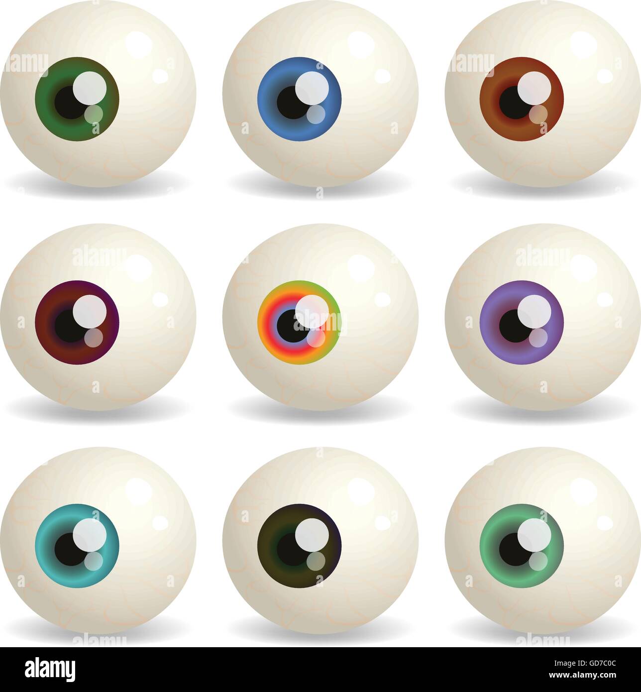
Human eyeballs Stock Vector Images Alamy

ma eye realistic human realtime Eyeball art, Eyeball drawing, Anatomy art
Eyeball

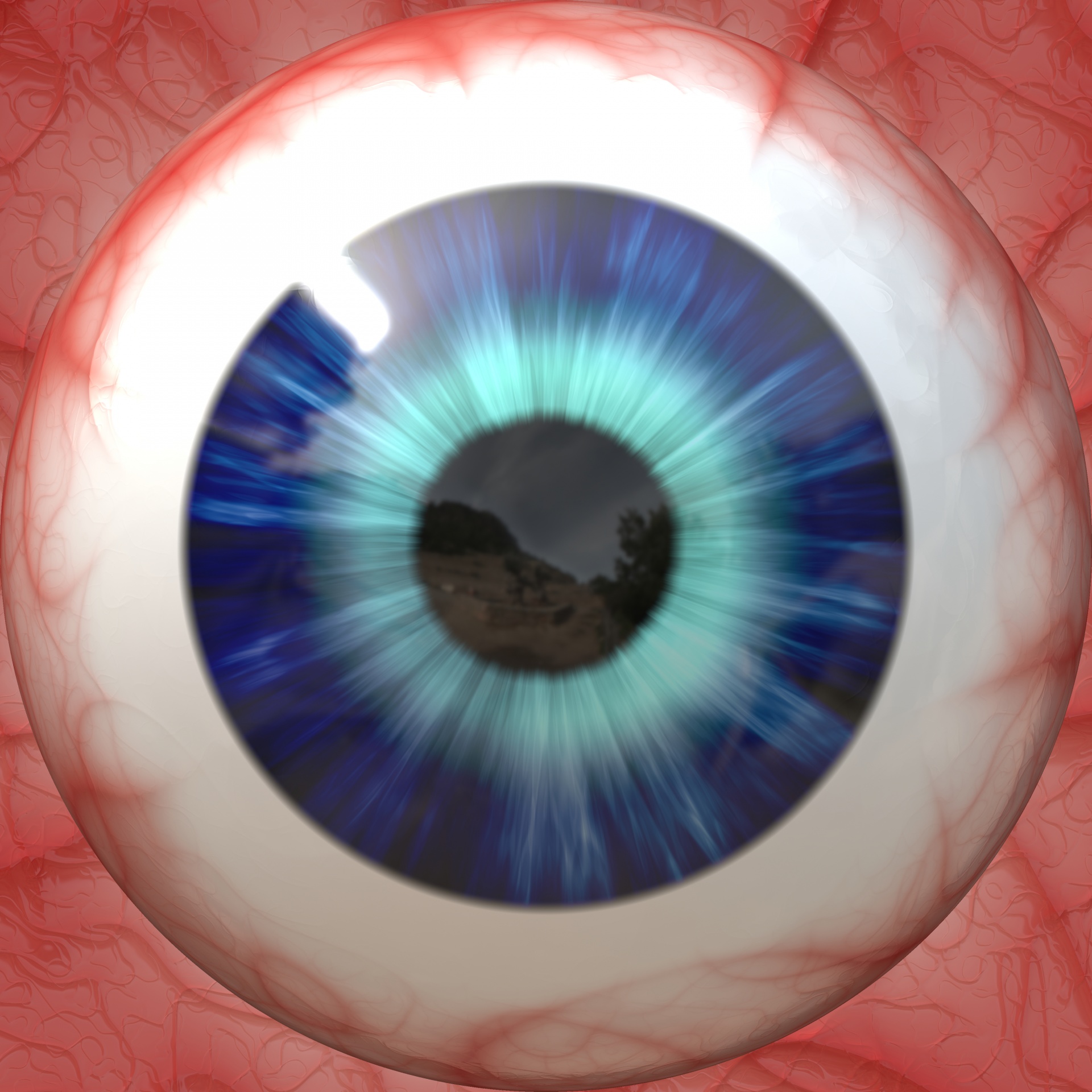
Premium Photo Human eye isolated on a white background realistic
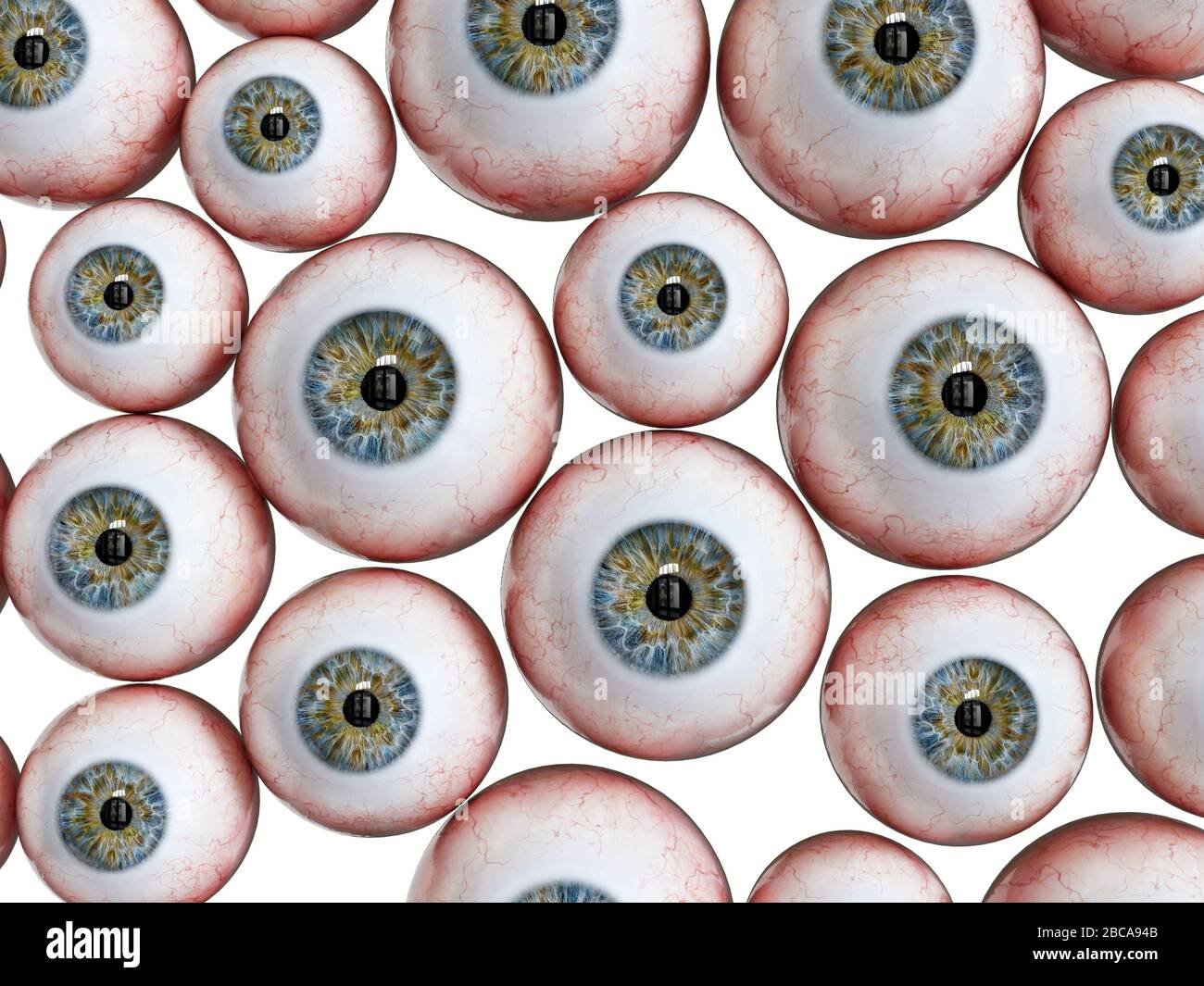
Eyeballs isolated hires stock photography and images Alamy

Two Realistic Human Eyeballs With Blue And Green Iris Isolated On White

Human eyeballs kesilkorean

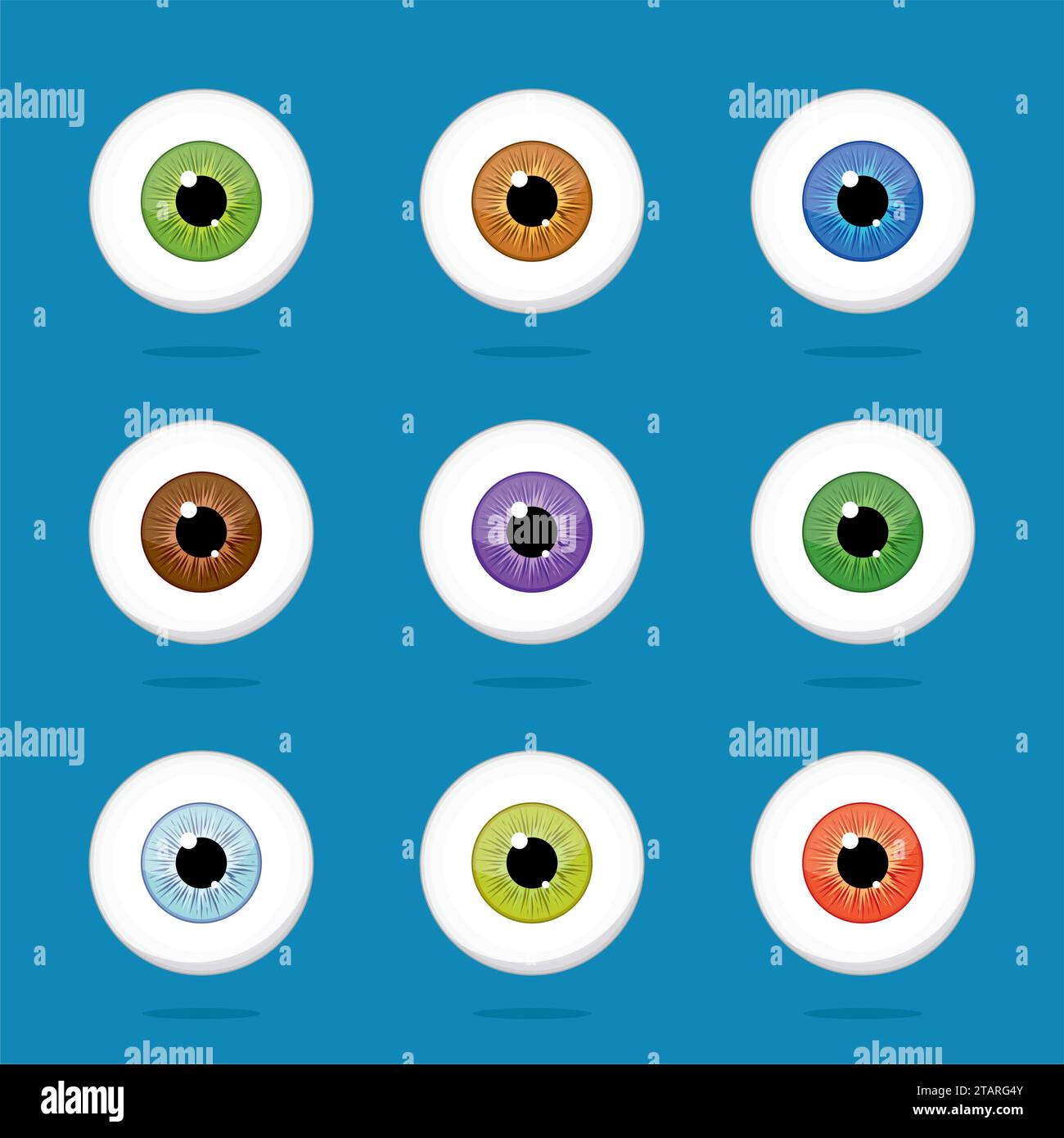
Human Eyeballs Iris Pupils Isolated On Blue Background Blue Yellow
Colorful human anatomy hires stock photography and images Alamy

Best Eyeball RoyaltyFree Images, Stock Photos & Pictures Shutterstock
The Meaning Of Eyeball Is The More Or Less Globular Capsule Of The Vertebrate Eye Formed By The Sclera And Cornea Together With Their Contained Structures.
These Remarkable Features Of Our Eye Are Enabled By The Complex Structure Of The Eyeball.
Buphthalmos Is An Eye Condition That’s Present At Birth.
Humans Have Two Eyes, Situated On The Left And The Right Of The Face.
Related Post: